Nikon Small World 2026 honors images of insects, neurons, algae and other tiny subjects, showing how microscopy can make hidden structures visible.
A closer look at familiar and unfamiliar forms
The 2026 Nikon Small World selections turn subjects that are difficult to see unaided into detailed visual studies. The recognized images range from weevils and water fleas to neurons, algae and the interior of a spruce treetop. Together, they show how microscopy can reveal structure and texture across very different kinds of specimens.
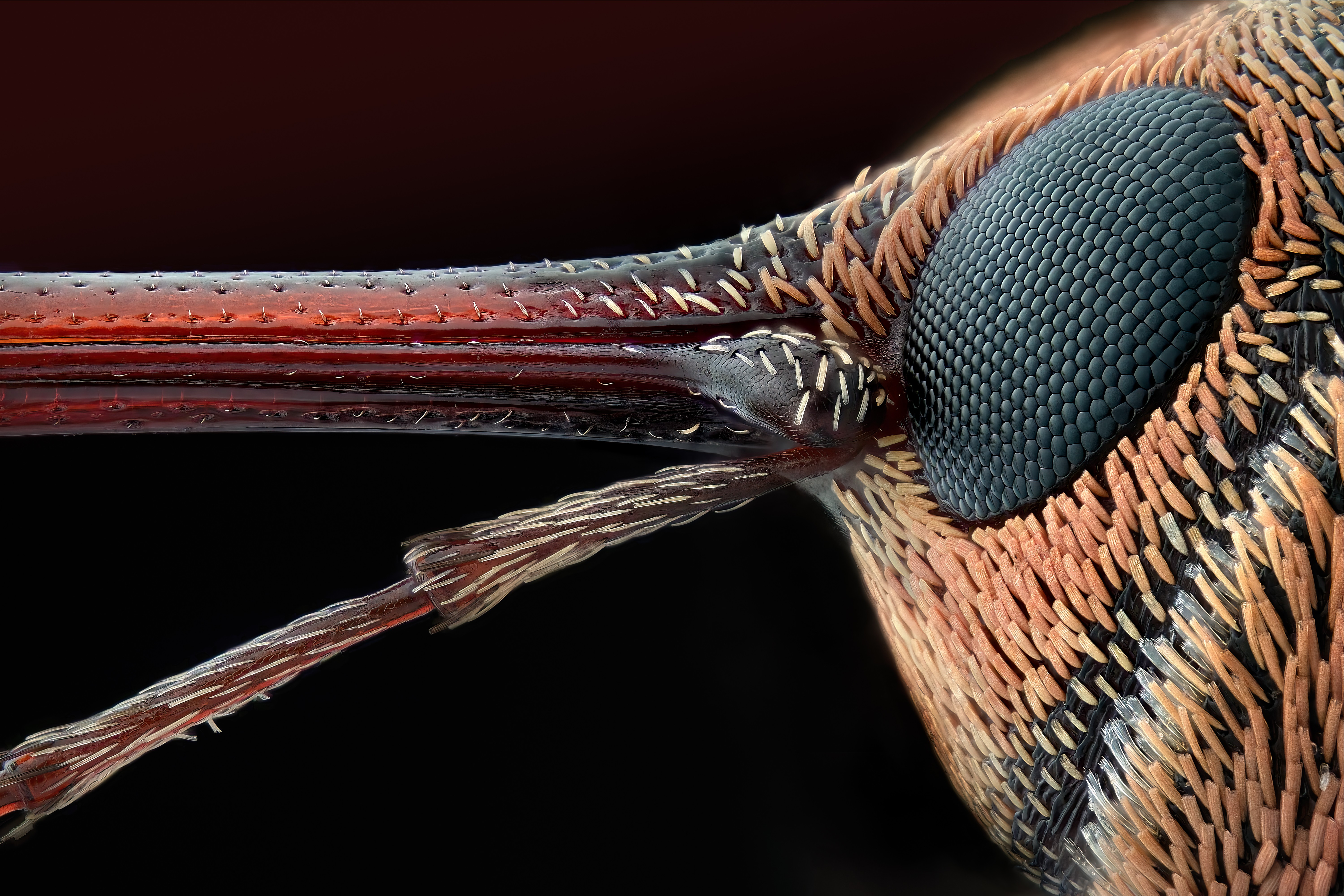
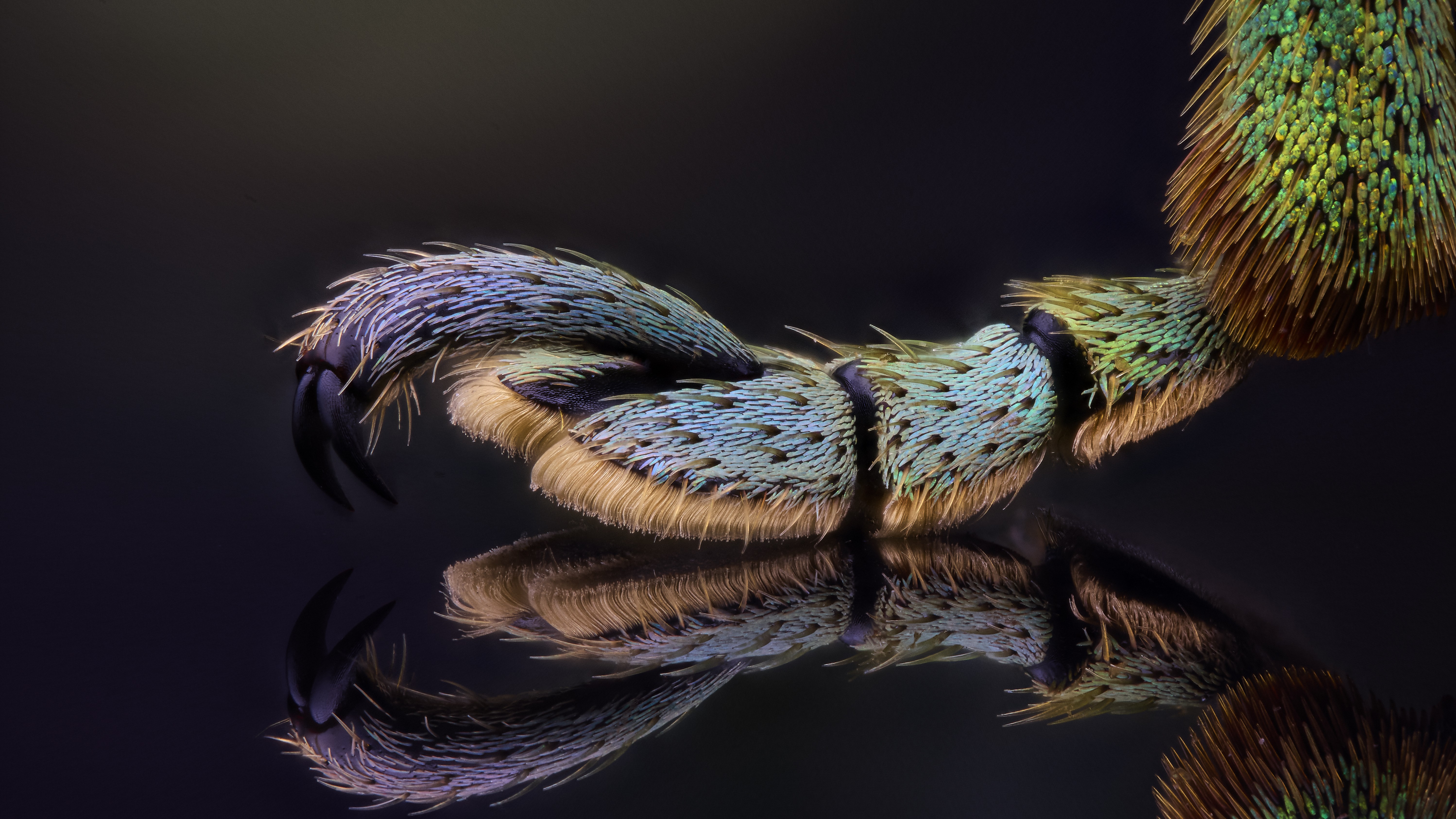
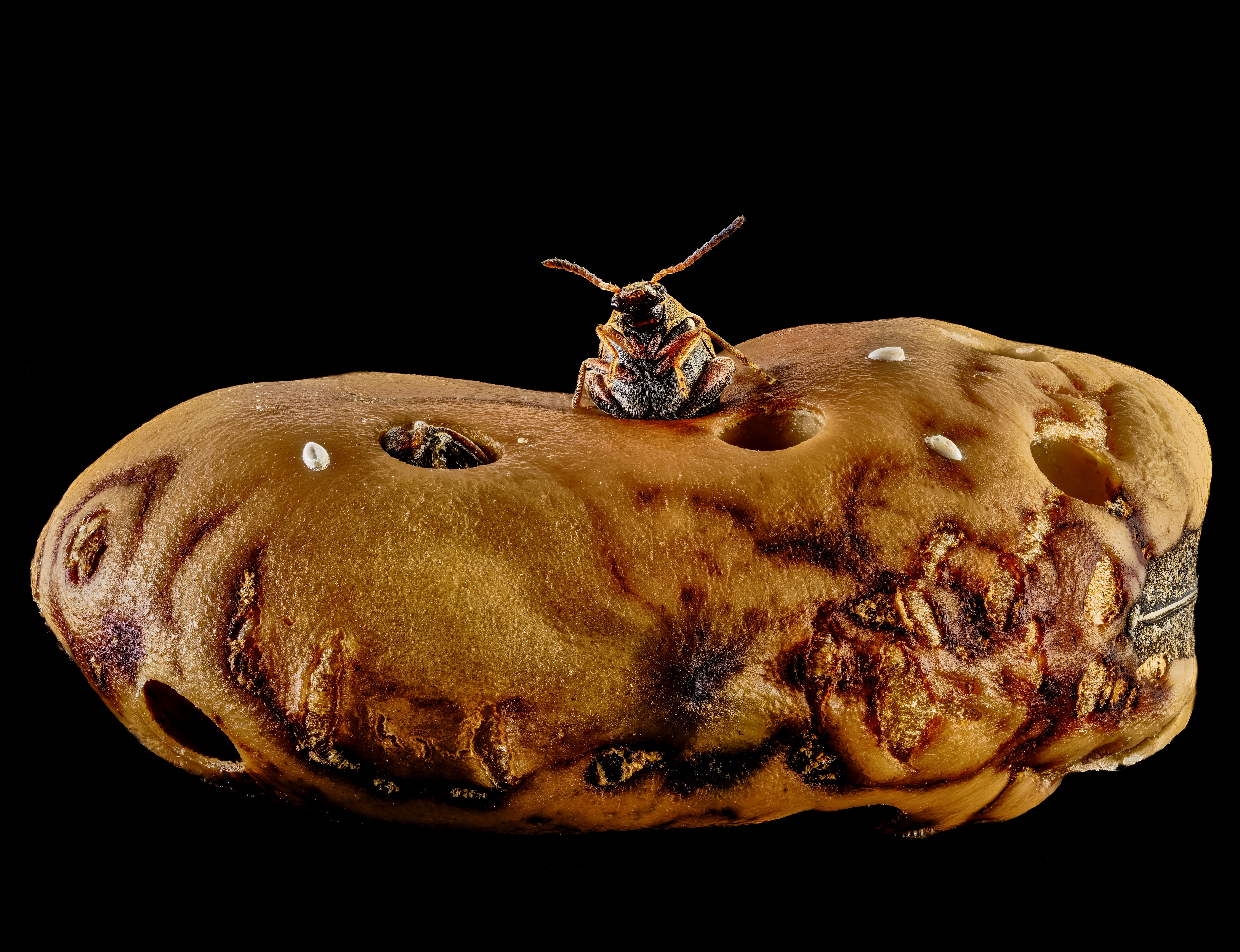
Several images focus on insects. Daniel Knop’s Image of Distinction isolates a weevil’s leg against a reflective surface, while Přemysl Orlík’s close view brings a hazelnut weevil’s face into focus. Zhang You captured a weevil emerging from a hole it bored into a fava bean. These views offer different ways into the same broad subject: a small animal’s body, its features and its relationship with the material around it.
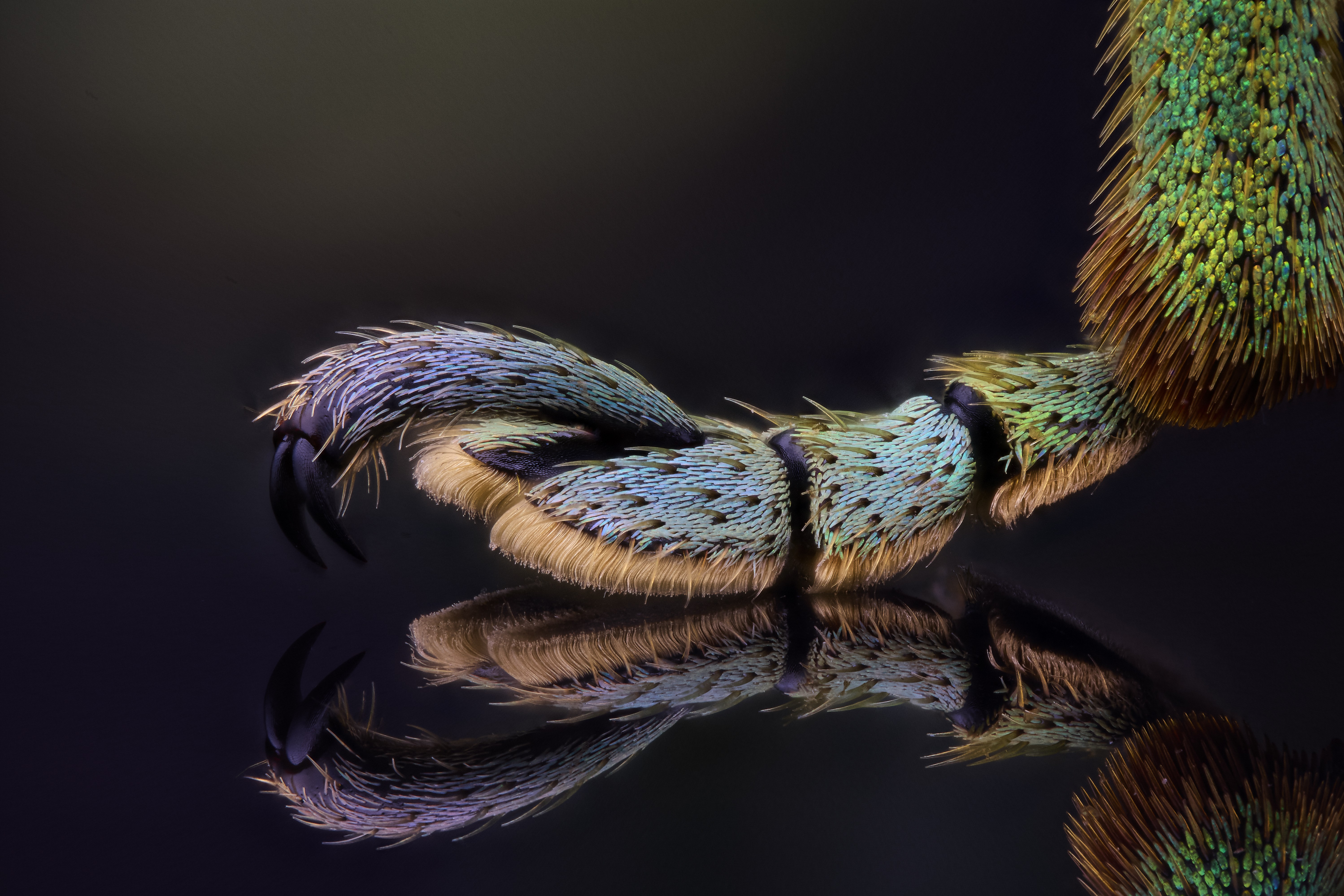
That variety matters to the viewing experience. A leg, a face and an insect at the entrance to a hole each direct attention to a different detail. Magnification makes those details visible, while lighting and composition help distinguish the subject from its background. The images are records of real specimens, but they also invite viewers to notice shapes and patterns they might otherwise overlook.
Living structures, from cells to whole organisms
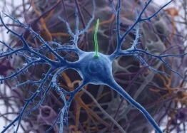
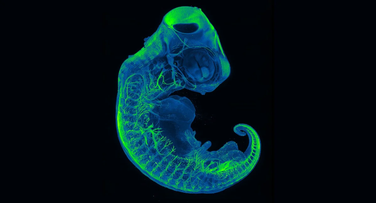
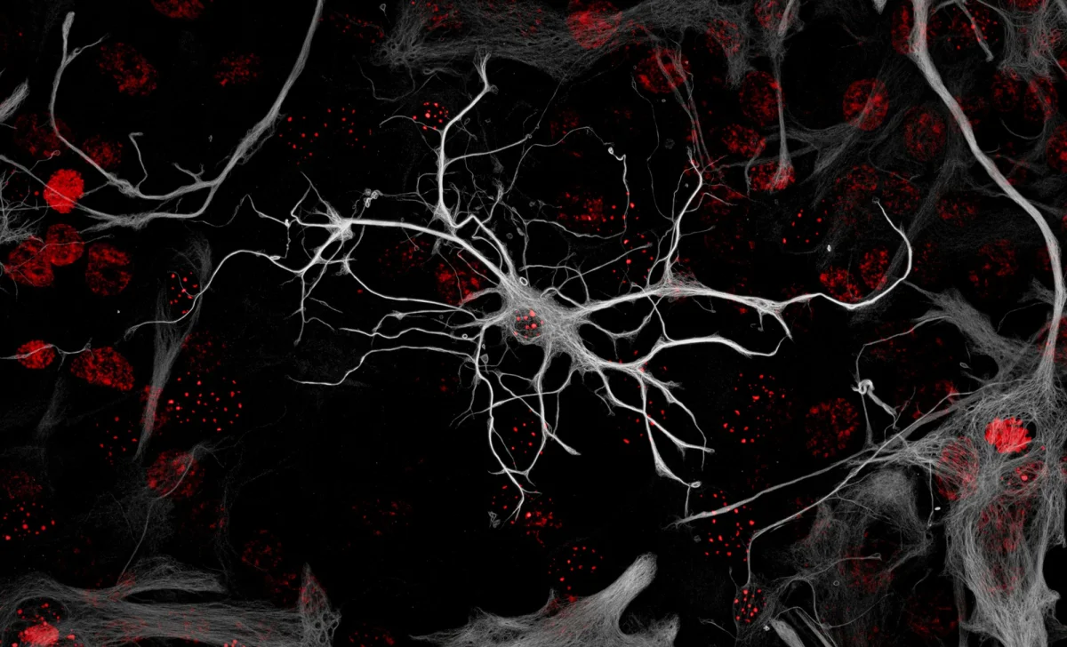
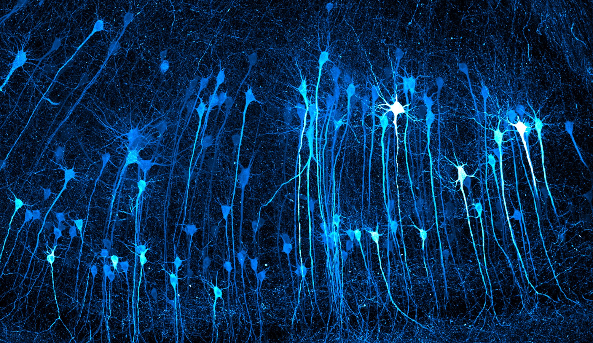
Other selections shift from the visible contours of insects to the organization of living tissue. Kaelin Wulf and Rachel Flannery of the Allen Institute received third place for an image of neurons in an expanded mouse brain. Nicolas Noel and Dr. Valentina Cigliola’s Image of Distinction shows a neonatal-mouse astrocyte in culture. An honorable mention went to a University of Cincinnati group for an image of neurons in a red-eared-slider turtle embryo.
These subjects are at different scales and stages of life. One image centers on neurons in an expanded brain, another on a single cultured cell, and another on neural pathways in an embryo. Their bright colors and fine branching forms can make complex biological structures easier to inspect visually. The pictures alone do not establish what those structures do or what they mean for health; their contribution here is to make parts of living systems visible in striking detail.
Small ecosystems and unexpected materials
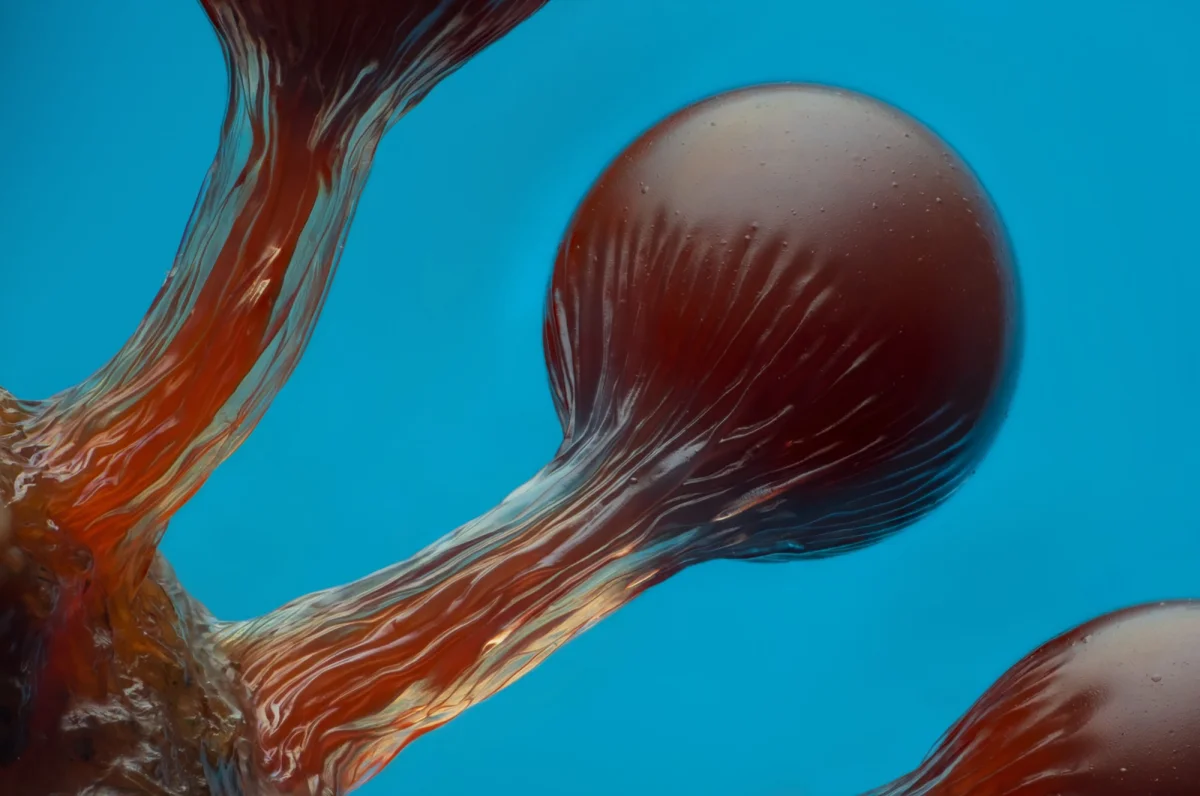
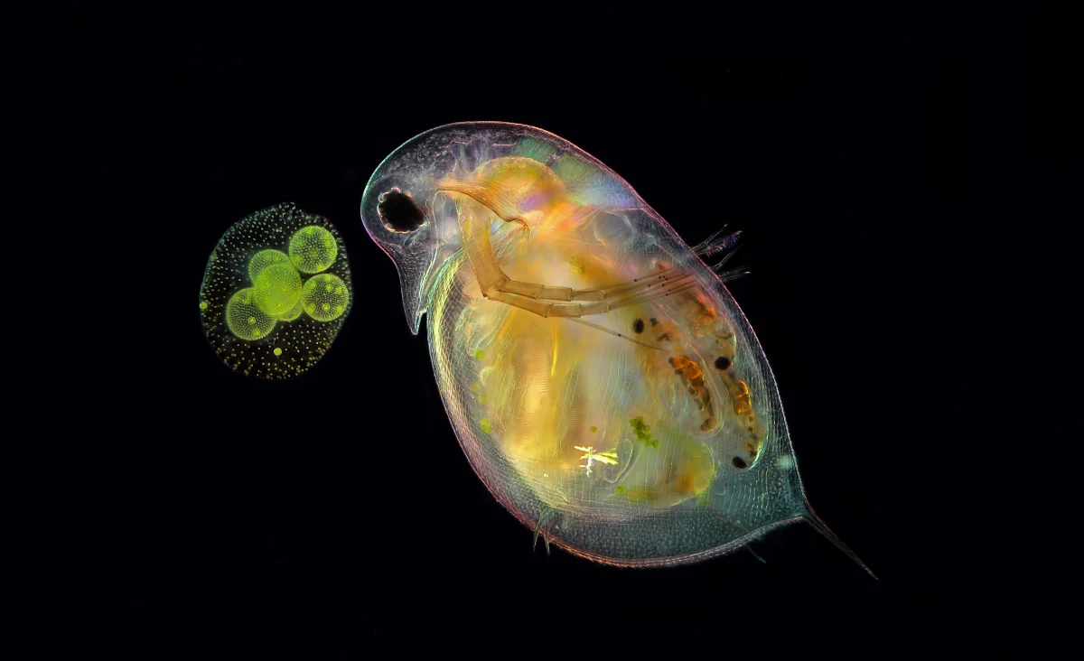
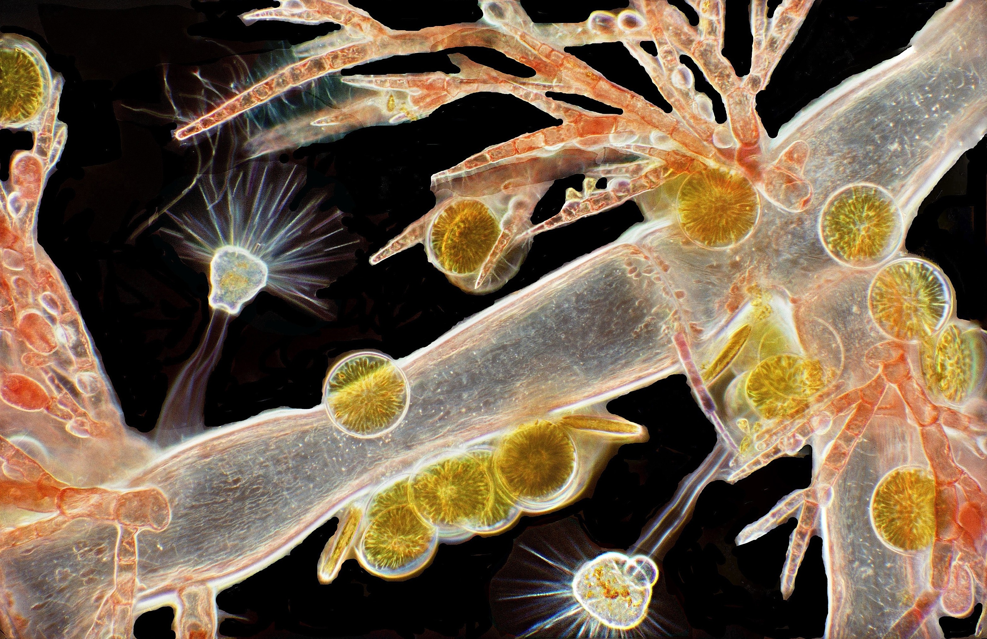
Some of the selected images place tiny organisms alongside one another. Anne Gleich’s Image of Distinction pairs a water flea, Daphnia, with green algae, Volvox. Dr. Arlene Wechezak’s image brings red algae, diatoms and protozoans into one view. Eric Epstein’s sixth-place image features the spore-bearing structures of a Hemitrichia slime mold, arranged as small spheres atop narrow stalks.
These scenes suggest how microscopy can reveal both individual forms and relationships within a sample. A water flea next to algae, or several microscopic groups in one frame, gives viewers more than a single isolated outline to study. The images do not describe the full ecology of these organisms, but they provide a close visual starting point for noticing their varied shapes and how many kinds of life can occupy a small field of view.
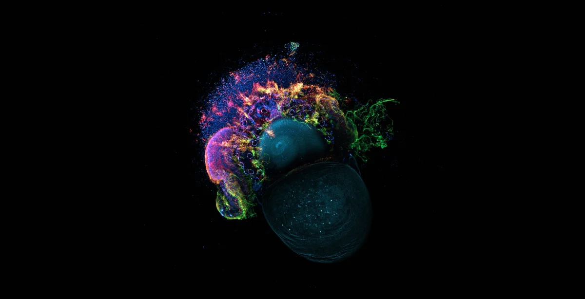
The collection also includes an image of a slipper-limpet larva, with its cellular structures illuminated in multiple colors. Another image shows an adult fungus gnat caught in the sticky trichomes of a cannabis plant. These choices widen the subject matter further: the microscopic view can show an organism’s internal arrangement, or the fine features of a plant that interact with insects.
Microscopy beyond biology
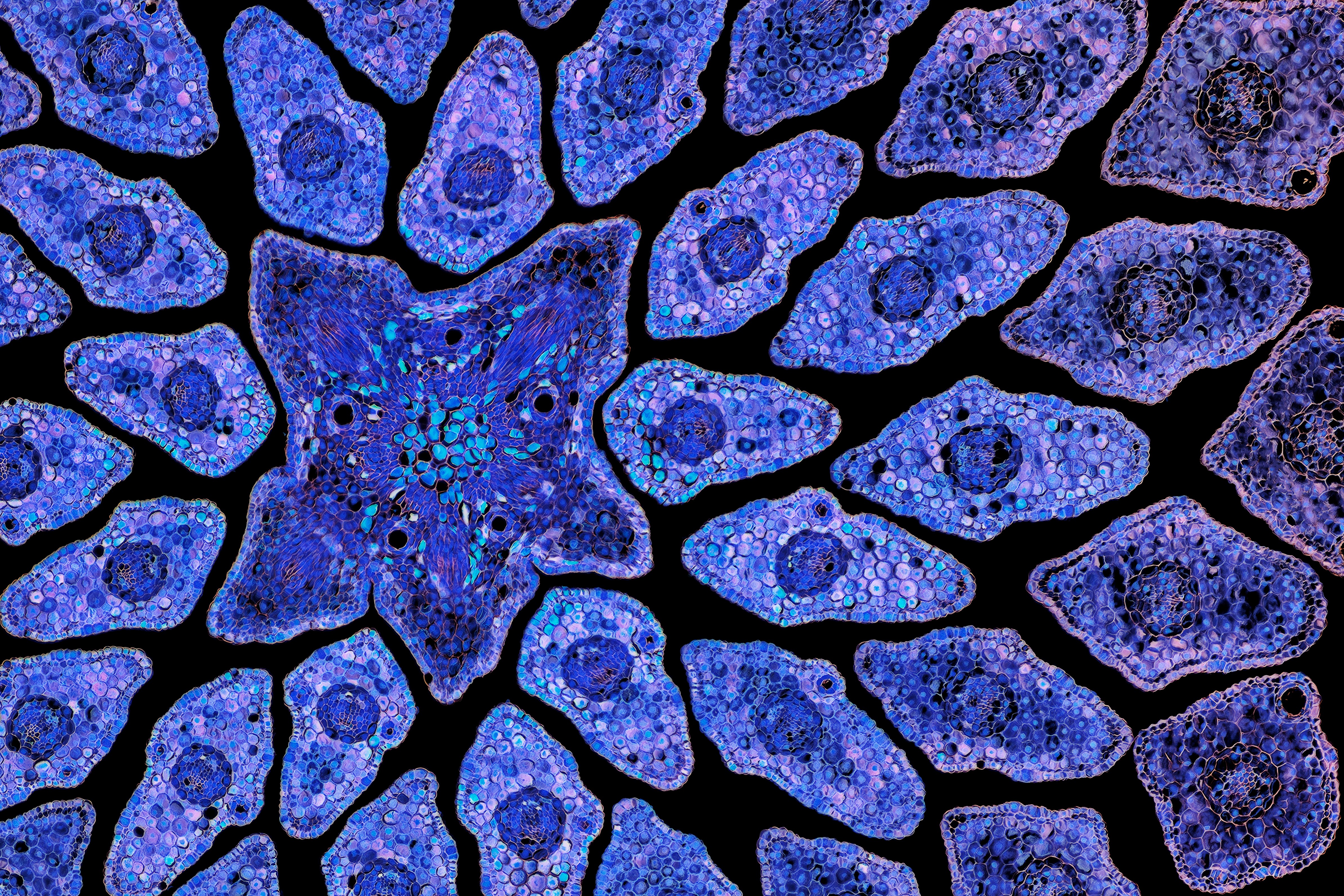
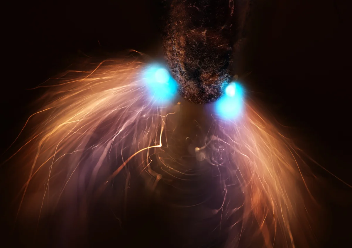
Not every selected subject is a living specimen. Dr. Marcel Clemens earned eighth place with a pencil lead photographed during exposure to a high-voltage electrical discharge. The image brings a familiar object into an unusual setting, where sparks and illumination become part of what the microscope records. Gerhard Vlcek’s spruce treetop cross-section offers another kind of material view, revealing purple forms within the tree’s structure.
The selections make a clear case for looking closely: a microscope can turn an insect, a cell, a plant cross-section or even a pencil tip into a subject with its own visual character. Viewers can find the full set of winning and honored images on the Nikon Small World website.