A cell study found that senescent cells can affect neighbors through vesicles, even when they release few familiar inflammatory signals.
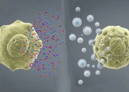
Senescent cells may influence their neighbors through more than the inflammatory substances commonly associated with cellular aging. In a laboratory study of human fibroblasts, cells made senescent by different routes released distinct mixtures of signals. Small extracellular vesicles from both groups affected recipient cells, even though one group released few of the familiar inflammatory factors. The findings suggest that the effects of senescent cells may depend on how they entered that state.

Two routes to a similar cellular state
Cellular senescence describes a state in which cells stop dividing. Senescent cells can also release a mixture of substances known as the senescence-associated secretory phenotype, or SASP. Its components can affect nearby cells. Inflammatory proteins such as cytokines are among the best-known components, but the mixture can also include metabolites and extracellular vesicles: small membrane-bound packages that carry cellular material.
The study, published in Aging, compared three groups of IMR-90 human fibroblasts: cells pushed into senescence by the cancer-related gene H-RASG12V, cells made to express p16, and non-senescent control cells. The p16 protein is encoded by CDKN2A and is widely used as a marker associated with senescence. Earlier research has linked p16-positive cells to aging, while laboratory work has also suggested that p16-driven senescence can have a different secretory profile from senescence induced by other routes.
That distinction matters for interpreting studies that use p16 as a marker. A marker can identify a feature of a cell without describing every process taking place inside it. The researchers’ comparison indicates that the route into senescence may shape what a cell releases, and therefore which effects it could have on surrounding cells.
Inflammatory signals differed between groups
The RAS-induced senescent cells released the inflammatory signal IL-1β. The p16-expressing cells and control cells did not release detectable amounts of it. The p16 group produced IL-6 messenger RNA, but the researchers found that it released less IL-6 protein into its surroundings than the control group.
The pattern for small extracellular vesicles (sEVs) was different. The controls released none detected in the experiment, the RAS group released many, and the p16 group released a smaller increase. This separation between vesicle release and familiar inflammatory signals is a key part of the report: low levels of some soluble inflammatory factors did not mean the p16 cells had no way to affect other cells.
These results come from cultured cells, so they do not establish how much either type of signal contributes to aging or disease in a person. The experiment is useful for comparing cellular behavior under controlled conditions, but a dish of fibroblasts cannot reproduce the mix of cell types and influences present in human tissue.
Vesicles carried effects to recipient cells
When the researchers exposed non-senescent cells to sEVs from the two senescent groups, the recipient cells showed different responses. Vesicles from p16-expressing cells increased p16 expression in recipient cells, but did not prompt them to release the inflammatory signals IL-6 and IL-8. Vesicles from RAS-induced cells did not consistently raise p16 expression, yet they did encourage production of those inflammatory cytokines.
Both types of vesicles increased DNA damage as measured by γH2AX, a biomarker used to assess a cellular response to DNA damage. The results suggest that vesicles from different senescent cells can produce distinct responses, while sharing at least one measured effect. They do not reveal which substances inside the vesicles caused those changes.
In a further experiment, vesicles derived from fibroblasts of older people were added to young fibroblasts in the laboratory. The recipient cells developed a senescence-associated signal measured by SA-β-gal. This supports the possibility that vesicles from older cells can affect younger cells under laboratory conditions. It does not show that vesicles alone drive senescence in living people, or that the same effect occurs in every tissue.
What the study leaves open
The authors describe the SASP as having a soluble component, made up of proteins and other smaller substances, and a vesicle component. In their experiment, p16-expressing cells released vesicles even though their familiar soluble inflammatory output was limited. This offers a reason to study both types of signals when assessing what senescent cells do.
There are important gaps. The researchers did not identify the specific contents of the vesicles or track changes in gene activity in recipient cells. They propose that vesicle cargo might damage DNA or trigger cell stress responses, but those explanations remain hypotheses. The experiments also do not show whether blocking vesicle release would improve health or alter aging in people.
For now, this is exploratory cell research, not evidence for a treatment or a reason to change care. It adds detail to how senescent cells may communicate, and it cautions against treating all senescent cells as if they send the same signals. The next step is to identify the active vesicle contents and determine whether these laboratory effects occur in living tissue.