In mice, blocking STING alongside NMN supplementation preserved bone measures, but the small study doesn’t show whether the approach benefits people.
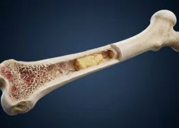
Aging bone marrow may weaken bone formation through a chain of signals linking marrow fat cells, immune cells and bone-forming cells. In a study of mice, researchers found that blocking the immune protein STING while supplementing NMN helped preserve bone density and structure. The findings offer a possible explanation for age-related bone decline, but they don’t establish that the approach works in people.
Immune signaling rose with age
Bone is continually remodeled: osteoclasts break down old tissue, while osteoblasts build new bone. Osteoblasts develop from bone marrow mesenchymal stem cells (BMSCs), which can also become fat cells. As marrow fat accumulates with age, the marrow tends to become more inflammatory and less supportive of bone formation. The reasons for this shift are still being studied.
The research team examined cGAS–STING, an immune signaling pathway that responds to DNA found in the wrong part of a cell. The enzyme cGAS detects this DNA and produces cGAMP, which activates STING and downstream inflammatory signals. The pathway normally helps cells respond to infections, but misplaced DNA, including DNA released from stressed mitochondria, can also trigger it.
In mice aged 3, 10, 18 and 24 months, the researchers observed progressively stronger cGAS–STING activity in bone marrow. They found more DNA in the cell cytoplasm, more cGAMP and increased activation of STING and related signaling proteins. Among the marrow cell types examined, macrophages had the highest baseline expression of the Sting gene, and their STING activity rose with age.
To test whether macrophage STING contributed to bone decline, the researchers used a viral vector to silence Sting in macrophages from 12 to 18 months of age. Treated mice retained more bone density and healthier bone structure, showed greater measures of new bone deposition, and had fewer marrow fat cells. These results support a role for macrophage STING in the process, though they come from an animal experiment.
Marrow fat cells may help sustain the signal
Experiments with cells pointed to communication between old macrophages and young BMSCs. Old macrophages made it harder for the stem cells to develop into bone-forming cells. Macrophages from which STING had been removed caused less harm, suggesting that this pathway contributes to the effect.
The team then investigated what might activate STING in aging marrow. Old mice had higher levels of endotoxin in their blood and marrow. Lipopolysaccharide (LPS), a component of bacterial membranes, activated STING in macrophages in the researchers’ experiments.
Signals from marrow fat cells offered another possible source. Secretions from old fat cells activated macrophage STING in laboratory experiments and after injection into the marrow of young mice. The researchers measured 44 proteins that were elevated in marrow with age and focused on DPP4, which fat cells released in greater amounts than the other marrow cell types tested. Old fat cells released more soluble DPP4 than young ones.
When researchers injected DPP4 into young mice’s marrow for four weeks, bone density and structure worsened and macrophage STING activity increased. Reducing Dpp4 expression in marrow fat cells improved one measure of bone structure: the spacing between trabeculae. Yet DPP4 alone didn’t significantly activate STING. It strengthened the response when combined with fluid from old marrow, suggesting it contributes to an inflammatory setting rather than acting as a sufficient trigger on its own.
The researchers found evidence that DPP4 can bind TLR4, a receptor involved in innate immune responses. Blocking TLR4 reduced STING activation. They also found more mitochondrial DNA outside mitochondria in old macrophages. Lowering mitochondrial DNA with low-dose ethidium bromide partly reduced STING activity, while other measures indicated greater mitochondrial stress in older cells. Taken together, the results suggest that signals from outside the macrophage and damage within it may work together to keep STING active.
NAD⁺ and the treatment question
Macrophage NAD⁺ levels declined with age in the study, while NAD⁺-degrading activity increased. Activating the pathway lowered NAD⁺; suppressing cGAS or STING countered the change. The researchers linked STING signaling to increased production of CD38, an enzyme that breaks down NAD⁺. They propose that CD38 on the cell surface may also reduce extracellular NAD⁺ and its precursor, NMN, limiting what nearby BMSCs can access.
Old BMSCs had poorer mitochondrial function and produced less ATP, the main energy-carrying molecule in cells. NMN supplementation partly improved these measures, although the improvement was smaller when BMSCs were exposed to old or artificially activated macrophages. Macrophages from old mice lacking Sting or Cd38 also supported better mitochondrial respiration.
In the final intervention, mice aged 12 to 18 months received the STING inhibitor H-151 together with NMN. The combination preserved bone density and structure, increased new bone deposition, reduced inflammatory markers, and improved NAD⁺ levels and BMSC mitochondrial function. The researchers compared the results with alendronate, an established osteoporosis medicine; bone structure improvements in the two groups were broadly similar.
The study was small, and because H-151 and NMN were given together, it can’t show how much each treatment contributed. It also tested mice, so it doesn’t establish a benefit or suitable treatment approach for people. The findings help map a possible connection between marrow fat, inflammation and bone formation. Human studies would be needed to determine whether that pathway is relevant to people managing age-related bone loss.